Type IV - delayed-type hypersensitivity
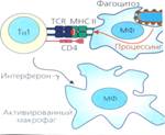
• immune tolerance (patience, unresponsiveness) - the body does not respond to hypertension and does not produce the AT. Occurs when the body met with hypertension in the embryonic period, for defects of lymphoid tissue, admission is very high or very low doses of antigen in an organism with a weak IP. • Immunological paralysis - the inability org ma-arr-ty AT when very large doses of antigen. Due to blockade of immunocompetent cells. After removing excess AG products AT vozobnavlyaetsya. • Immune deficiency - lack or loss of humoral and cellular defenses. M / b innate and acquired. Illustrative material:electronic version of multimedia lectures (the student receives at the Department), presented in the «Power Point» with photos, charts, tables, movies
Literature in Russian: Main: 1. Борисов Л.Б. Медицинская микробиология, вирусология, иммунология. - М.: МИА, 2001.- 734 с. 2. Табаева А.А. Микробиология поражений полости рта при стоматологических и инфекционных заболеваниях. Учебное пособие. – Алматы, 2006. -127с 3. Медициналық микробиология, Алматы, 2011.-683б Рамазанова Б.А, Кудайбергенұлы К.К редакциялаумен. 4. Ричард Дж.Ламонт., Роберт А.Берне., Мерилин С.Лантц., Дональд Дж.Лебланк., перевод с английского В.К. Леонтьева. Микробиология и иммунология для стоматологов. Практическая медицина. Москва 2010.- 502с. 5. Jacquelyn G Black “Microbiology”,7 th ,WILEY,2010,p.846 Test questions (feedback): 1. What are the main periods of the infectious process 2. What are the classification of infection? 3. Which factors pathogens znae6te you? 4. What is odontogenic infection? 5. Issdevaniya What methods used in microbiology? 6. How are odontogenic infections? 2 7. Which types of odontogenic infections isolated on the prevalence of the inflammatory process? 8. Describe certain types of odontogenic infections. 9. Which microorganisms can be agents of odontogenic infections? 10. Which types of immunity you know? 11. Name the cellular factors nonspecific defense 12. Name humoral factors nonspecific defense 13. What are the characteristics of immunity of the oral cavity? 14. What are the main cells of specific immunity? 15. Than antigen antibody differs from? 16. What cells produce immunoglobulins? 17. Immunoglobullinov What classes do you know? 18. Than primary immune response differs from the secondary? 19. How can manifest allergy to dental material? Тheme №3: "Pathogens of acute and chronic bacterial infections with lesions of the mouth and lips (coccal flora, pathogenic and opportunistic Corynebacterium, tuberculosis, leprosy, syphilis, actinomycosis)"; Objective:The main objective of the study of this topic is to develop students knowledge of coccal microflora and its role in the etiology of chronic inflammatory diseases, lesions of teeth and oral mucosa. Formation of knowledge about the spirochetes and actinomycetes causing defeat of the mouth and lips of the TB germs, leprosy and clinical features in lesions of the mouth and lips. Abstract of lecture: Stomatitis caused by streptococci (Fig. 1), and staphylococci (Fig. 8), constitute the main group of lesions. Stomatitis can be superficial and short-term or heavy unites the concept of "oral sepsis." In childhood watching impetigiozns stomatitis. For the disease characterized by the appearance on the mucosa of the lips, cheeks, gums, palate and tongue surface erosions, often merging together. Erosion covered with yellowish-gray patina, with its scraping bleeding occurs. Gums, especially at the free edge, often ulcerate. Originally isolated from the lesions streptococci (usually Streptococcus pyogenes), and at later - staphylococci (Staphylococcus aureus). Impetigo tends to spread purulent process in the skin (Fig. 11). Streptococcus pyogenes is also capable of causing erysipelas oral mucosa. The lesions may be an extension of inflammation on the skin of the face or start with small cracks and abrasions on mucous membranes of the mouth and pockets. Erysipelas sometimes develops after surgery and orthopedic surgery in the oral cavity. On the oral mucosa develops serous-hemorrhagic inflammation with marked edema. In the deeper layers of the mucosa develops leukocyte infiltration. Mucosa becomes dark crimson. In severe cases, it appears the bubbles and areas of necrosis. Local manifestations accompanied by symptoms of intoxication. In immunocompromised individuals possible generalization of the process with the development of sepsis. Another disease caused by streptococcus - Zayed (Fig. 12). The disease begins with the appearance of a small corner of his mouth streptococcal pustules, quickly transformed into scraps with erosion of the epidermis at the edges. In the absence of treatment and non-compliance with the basic rules of hygiene, and due to stretching of the skin upon expansion of the mouth and minor injuries in the middle of a crack erosion, turning on the buccal mucosa. Crack and bleed easily covered with bloody or purulent crust. Reinforced and drooling mouth raunchy content contribute to the continuous irritation streptococcal erosion, which can lead to streptococcal impetigo on the skin. Nonhemolytic streptococci included in the oral microbial biocenosis which constitute 30-60% of the microorganisms. This cariogenic streptococci biogroups S. mutans. They are distinguished by low virulence; they cause systemic involvement can be seen as opportunistic. The bulk of them constitute bacterial endocarditis developing after penetration of bacteria into the bloodstream when traumatizing mucous membranes (eg, after brushing your teeth, chewing roughage). Endocarditis are malignant in nature and are accompanied by damage to the heart valves. Ability to cause endocarditis caused by structural features of glycans (dextran) cell walls of bacteria, Streptococcus facilitate adhesion to aggregates of platelets and fibrin on the damaged valves. Opportunistic Corynebacterium C. xerosi, C.pseudodiphtheriticum and others are normal inhabitants of the skin and mucous membranes of the mouth, eyes and genitals. C.diphtheriae causes diphtheria, an acute infectious disease, which is characterized by specific inflammation throat, larynx, trachea, or fibrinous dr.organov to form films and intoxication. (Diphtera - film). Refers to the number of infections toksinemicheskih (exotoxin circulates in the blood). Diphtheria - gram + bacillus, motionless, with volyutinovymi grains on the ends. Ill mostly children, especially toddlers and kindergarten age. Exotoxin first strikes directly the epithelial cells, and then the nearby blood vessels, increasing their permeability. In exiting the vascular exudate found fibrinogen, which forms on the surface of clotting grayish - white filmy raids tightly welded with the surrounding tissue. They are taken seriously, when they divorced exposed erosive surface. Proliferation of these films and their transition to the airways lead to the development of true croup and asphyxia. TUBERCULOSIS - chronic infection manifested specific visceral organs and tissues. Pathogen R.Kohom opened in 1882 Tuberculosis - affects the respiratory system. This is a very real issue. About 8 million people become ill with tuberculosis in the past year. About 50 million people may be infected with mycobacteria resistant to drugs. Every patient with active TB disease during infect an average of 10-15 people. Pathogen refers to the order Actinomycetales, family Mycobacteriaceae, the genus Mycobacterium. Greatest importance in human pathology play types: M.tuberculosis, M.bovis, M.avium, M.africanum. Pathogen relates to acid-resistant bacteria, as cell wall contains a lot (up to 60%), lipids, waxes and phosphatides. So badly stained with aniline dyes. Ziehl-Neelsen stained red. Lesions in the mouth of tuberculosis: primary - tubercular ulcers in children; secondary - lupus, ulcerative miliary tuberculosis, tuberculous gumma, erythema indurativny Leprae (leprosy, Hansen's disease) - a chronic generalized infection with primary defeat of derivatives of the ectoderm (skin and nervous system). Pathogen opened in 1873 G.Hansenom. refers to the order Actinomycetales, family Mycobacteriaceae, the genus Mycobacterium, an M. leprae. Disease manifests itself in two basic forms: tuberculoid form Har-Xia overgrowth of granulation tissue in the skin and mucous membranes with severe anesthesia. Relatively benign. Lepromatous form heavier. Observed reddish brown confluent infiltrates without anesthesia, loss of eyebrows and eyelashes, develops "lion face". In the mouth infiltrates found on the soft and hard palate, lips, tongue, as well as mucous along the nerve trunks. Lepromatous nodes may break, emitting a large number of mycobacteria. Mucosal lesions usually combined with skin lesions. Actinomycosis person observed in 55-60% of all patients with actinomycosis (thoracic, abdominal, actinomycosis of the genitourinary system, bones, central nervous system, generalized) and 6-10% of people with inflammatory lesions of the jaws and face. The main causative agents of actinomycosis - Actinomyces israelii and Actinomyces viscosus (Fig. 4). These actinomycete inhabit the oral cavity as saprophytes, in particular carious dental cavity in tartar deposits. Pathogenic potential of microorganisms is very low and actinomycoses develop only due to lower resistance due to vitamin deficiency, severe illness, etc. The most frequently observed actinomycosis neck-face area and the mandible (Fig. 13). Pathogen overcomes the epithelial barrier of the oral mucosa in trauma, surgery, injections. In the mucosa or in the deep soft tissues of developing one, and often several dense nodes granulomas (aktinomikomy) without acute inflammation, fever and health violations. Signs of intoxication with headaches, general weakness and low grade body temperature appear only in the decay of nodes with pus through several narrow fistulas. With the localization of nodes in the lower jaw often develops convulsive spasm of the muscles of the mouth (lockjaw), complicating meal. In the merger of several nodes has a significant inflammatory infiltrate density, which is an important diagnostic feature. In the center of the infiltrate formed several holes representing bulging red ("flesh color") as a nipple. Of fistulas stands liquid pus rich in yellowish-gray grains of an average of 0.3-2 mm, the so-called "sulfur granules" (calf Bollinger). Grain - Druze clusters formed mycelium clavate peripheral swellings. When detected, the diagnosis is obvious. The disease runs a chronic but often complicated by secondary bacterial infections; possible damage to the skin, muscles, glands, tongue, salivary glands and bone. Fuzospirohetoz - is an acute inflammation of the gums with pronounced symptoms of alteration. The etiology and pathogenesis of this disease is not fully disclosed. Necrotizing ulcerative gingivitis developing against catarrh. This disease occurs mainly in young adults, as a result of SARS, sore throat, flu, hypothermia, stress, malnutrition, hypovitaminosis reduced immunity. Against this background, the conditions for increasing the amount of microflora and increase its pathogenicity. Among the anaerobic microorganisms predominate form - fuzobakterii (Fusobacterium plautii), spirochetes (Treponema vincentii). Often precedes the development of the disease the inflammation caused by staphylococci and streptococci. Pathogenetic value fusiform bacteria associated with the presence of collagenase type tissue-dissolving enzymes, proteases, hyaluronidase, which cause the degradation of connective tissue. While low molecular weight nitrogen-containing products formed from the collapse of collagen, can be assimilated by spirochetes. Anaerobic conditions created in the necrotic tissue oxygen due to the action of inactivating bacterial enzymes catalase and superoxide dismutase, impede rapid recovery and contribute to further tissue damage. As a result, significant necrosis of the gums, the deformation of the gingival margin and creates a potential hotbed of chronic inflammation in the periodontium. Furthermore, during the life of secreted bacterial fatty acids, as well tissue-dissolving enzyme and inhibit Ig A protease immunity of mucous membranes of the mouth, causing the degradation of immunoglobulins complement. Together with fusiform bacteria in the inflammation develops other anaerobes: Bacteroides, peptokokki, peptostreptokokki, veylonelly. Development of necrotizing gingivitis Vincent promotes bad individual oral hygiene, dental plaque, the presence of caries, decayed teeth, shortness eruption of wisdom teeth (the presence of "the hood"). The patient complains of pain in the gums, which makes it difficult eating and talking. The examination determined hyperemia, presence of ulcers and necrosis gums (gray patina), a significant amount of soft plaque and hard dental plaque, there is halitosis. Often observed loss of the tonsils and the larynx with the development of a condition known as angina Simanovskiy-Vincent-Plaut. SYPHILIS - chronic venereal disease with cyclic course. Pathogen open F.Shaudinym and E. Hoffman in 1905, belongs to a Pathogen: Spirochaetales, family Spirochaetaceae, genus Treponema, Treponema pallidum mind. This spiral bacteria having 8-14 curls, identical in height and width, mobile, poorly painted anilinivymi dyes. Romanovsky-Giemsa stain in pale pink Lesions in the oral cavity with primary syphilis - chancre. Lesions in the oral cavity of secondary syphilis: papules and roseola located on the lips, tongue, bow, tonsils, on the buccal mucosa - on the line between the teeth at the tip and lateral surface of the tongue, tonsils - syphilitic angina. Lesions in the oral cavity with tertiary syphilis - syphilis Cuspal gummy. Lesions in the oral cavity of congenital syphilis: Infants syphilitic rash - 30%. Syphilides as papules as secondary syphilis. Syphilitic diffuse infiltration of the skin on the palms, soles, buttocks ivokrug mouth perioral scars, vermilion border, mucosal skin and cheeks. Linear scars in the corners of the mouth (scars Robinson-Fournier) Illustrative materials: Presentation in «PowerPoint» with photos, charts, tables, movies Literature in russian: Main: 1. Борисов Л.Б. Медицинская микробиология, вирусология, иммунология. - М.: МИА, 2001.- 734 с. 2. Табаева А.А. Микробиология поражений полости рта при стоматологических и инфекционных заболеваниях. Учебное пособие. – Алматы, 2006. -127с 3. Медициналық микробиология, Алматы, 2011.-683б Рамазанова Б.А, Кудайбергенұлы К.К редакциялаумен. 4. Ричард Дж.Ламонт., Роберт А.Берне., Мерилин С.Лантц., Дональд Дж.Лебланк., перевод с английского В.К. Леонтьева. Микробиология и иммунология для стоматологов. Практическая медицина. Москва 2010.- 502с. 5. Jacquelyn G Black “Microbiology”,7 th ,WILEY,2010,p.846 Additional: 1. Воробъев А.А., Кривошеин, Широбоков В.П. Медицинская и санитарная микробиология. – М.: Издательский центр «Академия», 2003. – 464 с. 2. Коротяев А.И, Бабичев С.Л. Медицинская микробиология, иммунология и вирусология. - СПб.: Спец.лит, 2000.- 591 с. 3. Тец В.В. Руководство к практическим занятиям по медицинской микробиологии, вирусологии и иммунологии. – М., 2002 4. Компьютерная программа “Диаморф” - “Медицинская микробиология” - атлас-руководство по бактериологии микологии, протозоологии и вирусологии под редакцией акад. проф.Воробьева А.А. Test questions (feedback): 1. Which gram-positive cocci can cause chronic inflammatory diseases in humans? 2. Which gram-positive cocci play a role in the occurrence of dental caries? 3. Which diseases are caused by gram-negative coccal flora? 4. What morphological features of causative agent of syphilis? 5. What are the manifestations of primary syphilis of the mouth and lips? 6. Clinical features of secondary syphilis of the mouth and lips. 7. What safety rules must comply with the dentist for the prevention of occupational infection with syphilis? 8. Actinomycosis infection What are the ways? 9. Name morphological and cultural characteristics of Mycobacterium tuberculosis? 10. Are there specific drugs for the prevention of tuberculosis? Leprosy? What? 11. How to control the state of immunity in tuberculosis? 12. Which method of research is used in the diagnosis of leprosy? 13. Which clinical manifestations of tuberculosis and leprosy in the mouth? 14. Which staining methods used for detection volyutinovyh grains in diphtheria? 15. Toxigenicity What causes strains of diphtheria? 16. Which manifestations of diphtheria in the mouth? Тheme №4 "Pathogens viral infections with lesions of the mouth and lips. Orthomyxoviruses. Paramyxoviruses. Adenoviruses. Picornaviruses. HIV. HSV. Rhabdoviridae. papovaviruses "; Objective:The main objective of the study of this topic is to develop students knowledge of lesions of the mouth and lips caused ortomiksovirusov, paramyxoviruses, adenoviruses, picornaviruses. Assimilation of knowledge about the epidemiology, biological properties of HIV, clinical manifestations of HIV infection and AIDS in the mouth. Formation of students' knowledge about the biological properties of herpes viruses, rhabdoviruses, papovaviruses and the role of these pathogens in the etiology of dental diseases. Abstract of lecture: The family belongs ortomiksovirus is influenza virus. (orthos - right, myxo - mucus.) This is a complex containing RNA virus circular shape with a diameter of 80 - 120 nm. Superkapsid contains spicules - spinous process of the neuraminidase (N) and hemagglutinin (H). This major surface Ag virus. Influenza virus type A is divided into 13 antigenic subtypes of hemagglutinin and by 10 to neuraminidase. Cause human disease subtypes H1, H2, H3 and N1, N2. About every 10 years, flu pandemics take on the character - this is due to the change of H and N-Ar-s virus type A (Ar drift and shift)-1918 "Spanish flu» (NSW1); 1957, "Asian" flu (N2N2); 1968, "Hong Kong" (H3N2). Influenza viruses B and C have a higher stability of Ag: V - is less intense and localized outbreaks of the epidemic; C is the cause of sporadic outbreaks; And - very volatile, so is the greatest danger of an epidemic. Source of infection - patients and virus carriers. Transmission - by airborne droplets (sneezing, coughing). Increase in the incidence observed in the colder months. Used for the prevention of influenza vaccines that induce humoral immunity and very slightly - cell. Immunity short, so annual vaccination is required. The main reason for the lack of effectiveness of influenza vaccines is the high variability of the circulating influenza virus, the emergence of a new subtype of the virus. To paramyxovirus pathogens include parainfluenza, mumps, measles, RSV infection. It is an infection with airborne transmission, the source of which serve patients and virus carriers. HPIV (human parainfluenza virus) has 5 serotypes. Causes SARS, colds, laryngotracheobronchitis, bronchiolitis and pneumonia. Often there are epidemic. Specific prophylaxis is not applicable. Mumps - infection characterized by a primary lesion of the parotid glands and the ability to cause outbreaks. There is one serotype of mumps virus. For specific prophylaxis used live vaccine A.A.Smorodintseva, mono-or associated with measles. For later use prevention and treatment specific immunoglobulin. RSV - respiratory syncytial virus infects most children during the first 6 months., Causes severe bronchitis, pneumonia. RS virus has immunosuppressive properties, so frequent secondary bacterial complications. Often causes recurrent disease, since immunity lasts no more than 1 year. RS virus common cause of nosocomial infections (pneumonia in infants and young children). Specific prophylaxis is not applicable. Adenoviruses - isolated in 1953 from the adenoids. Vozbuditkl - DNA - containing a virus belonging to the family Adenoviridae. Source of infection - patients with acute or latent form. Routes of transmission: airborne and fecal-oral. Most sick children from 6 months. up to 2 years. Manifested in the form of respiratory disease, pneumonia, conjunctivitis, gastroenteritis. Sometimes there is a shift in the chronic form (hr.tonzillity, sinusitis, tonsillitis). Possible allergy (asthmatic bronchitis and laryngotracheitis). Specific prevention: live adenovirus vaccine (United States also apply - leukocyte interferon deoxyribonuclease. Picornaviruses - is small (about 28 nm) Simple RNA containing viruses are capable of replication in the intestine and associated lymphoid tissue of the nasopharynx. The composition of the family Picornaviridae includes 4 genera: Enterovirus (poliovirus, coxsackievirus groups A and B, ECHO virus, hepatitis A); Rhinovirus -; Aphtovirus - FMD virus; Cardiovirus - rare in humans, mainly in animals. Poliomyelitis - an acute infectious disease characterized by lesions of the anterior horns of the gray matter of the spinal cord, and therefore develop lifelong limp paralysis of the lower limbs. Most sick children. For specific prophylaxis use oral and parenteral polio vaccine. A Coxsackie enteroviruses cause gerpanginy that appear vesicular eruptions on the back of the throat, dysphagia, anorexia. Vesicles burst to form the AFL with whitish down. The disease lasts 7-10 days and goes away without treatment. In dentistry are important aftovirusy causing foot and mouth disease and vesicular stomatitis. Human immunodeficiency virus belongs to the family Retroviridae, genus Lentivirus. This RNA virus contains a spherical shape (100-120 nm). The HIV pandemic swept the globe and is steadily spreading among people, carrying a deadly threat. Since the first HIV infected registering only 65 million people, 23 million people died. Registered in Kazakhstan about nine thousand HIV - infected. The virus infects cells that have CD 4 receptor - it lymphocytes, monocytes and macrophages. HIV replication leads to the death of T-helper lymphocytes, which weakens the immune system. HIV has a high variability that occurs in the course of infection and virus carriers in the body. This enables the virus to evade specific antibodies and cellular immune factors that leads to the chronic process. HIV infection - a slowly progressive infectious disease that affects the immune system, causing the body becomes susceptible to opportunistic infections and tumors, which eventually leads to the death of the patient Material for the study of HIV infection - blood, peripheral blood leukocytes, punctate l / nodes and bone marrow, genital mucosal secretions, semen, saliva, tear fluid, breast milk, cerebrospinal fluid, cadaveric material. Methods of diagnosis: serum (Immuno fluorescence analysis, immunoblot), genetic (polymerase chain reaction, genotyping), immunological (quantitative determination of CD4 and CD8 cells), virological (viral culture on leukocytes from healthy donors) There are no drugs, depleting the virus from infected cells, but antiretroviral therapy designed to affect different stages of viral replication. Specific prevention has been developed. By preventing HIV infection in the workplace include universal precautions: ispolozovat personal protective equipment (gloves, goggles, mask, all patients considered as potentially infectious. The clinical manifestations of HIV infection in the mouth include: recurrent aphthous stomatitis, angular cheilitis, recurrent herpes, oral candidiasis (thrush), hairy leukoplakia. Herpes - this DNA-containing complex viruses are subdivided into subfamilies: b-herpesviruses (herpes simplex virus, varicella zoster); in-herpesviruses (cytomegalovirus, HS 6 and 7 type (rash, chronic fatigue syndrome)); r-herpesviruses (Epstein-Barr virus (Burkitt's lymphoma, Kaposi's sarcoma, carcinoma of the nasopharynx)). Ubiquitous. Reservoir - an infected person. Lesions in the mouth often occur on the lips, buccal mucosa may be on, tongue, pharynx, tonsils. First, there is a feeling of burning, stinging, redness and swelling then, then there are a few small rounded painful vesicles with turbid fluid. Vesicles may become pustules and form of erosion. The patient is observed malaise, dysphagia, fever, regional l / y increase and painful. The causative agent of rabies belongs to the family Rhabdoviridae, genus Lyssavirus. Rabies - acute zoonotic infection, causing human and animal deaths irreversible neuronal damage central nervous system. Virion bullet shaped, helical nucleocapsid symmetry type in - single-stranded RNA. Reservoir of infection - warm-blooded animals dogs, cats, foxes, wolves, jackals, bats, etc. Transmitted through the bite or oslyunenie. Man - a biological dead end. In neurons, brain and spinal cord occurs intensive reproduction of the virus to form cytoplasmic inclusions Babes - Negri. Used for the prevention of rabies vaccine of the Fermi type of fixed inactivated virus, rabies gamma globulin, inactivated culture vaccine. Effective treatments exist. Infection always leads to death. Symptomatic therapy to reduce the suffering of the patient. Papovaviruses - human papilloma viruses. Papilomavirusnaya DNA integrates into the cell genome. Epithelial cells divide, forming a tumor growths, reaching terminal differentiation, stop dividing, they accumulate keratin. Genotype determines the location of the cancer. Genital warts are formed, oral, flat warts, genital warts, papilloma oral mucosa, conjunctival papillomas. Tumors can ozlokachestvlyatsya, metastasize. Surgical treatment. Illustrative materials: Presentation in «PowerPoint» with photos, charts, tables, movies Literature in Russion: Main: 1. Борисов Л.Б. Медицинская микробиология, вирусология, иммунология. - М.: МИА, 2001.- 734 с. 2. Табаева А.А. Микробиология поражений полости рта при стоматологических и инфекционных заболеваниях. Учебное пособие. – Алматы, 2006. -127с 3. Медициналық микробиология, Алматы, 2011.-683б Рамазанова Б.А, Кудайбергенұлы К.К редакциялаумен. 4. Ричард Дж.Ламонт., Роберт А.Берне., Мерилин С.Лантц., Дональд Дж.Лебланк., перевод с английского В.К. Леонтьева. Микробиология и иммунология для стоматологов. Практическая медицина. Москва 2010.- 502с. 5. Jacquelyn G Black “Microbiology”,7 th ,WILEY,2010,p.846 Additional: 1. Воробъев А.А., Кривошеин, Широбоков В.П. Медицинская и санитарная микробиология. – М.: Издательский центр «Академия», 2003. – 464 с. 2. Коротяев А.И, Бабичев С.Л. Медицинская микробиология, иммунология и вирусология. - СПб.: Спец.лит, 2000.- 591 с. 3. Тец В.В. Руководство к практическим занятиям по медицинской микробиологии, вирусологии и иммунологии. – М., 2002 4. Компьютерная программа “Диаморф” - “Медицинская микробиология” - атлас-руководство по бактериологии микологии, протозоологии и вирусологии под редакцией акад. проф.Воробьева А.А. Test questions (feedback): 1. Oral lesions caused by orthomyxoviruses. 2. Infection by ortomiksovirusov, paramyxoviruses, adenoviruses? 3. Which clinical symptoms observed in FMD in the mouth? 4. What are the characteristics of HIV replication? 5. Why is there no effective vaccine for AIDS? 6. AIDS-related opportunistic infection in the oral cavity. 7. What safety rules must comply dentist to avoid iatrogenic autoinfection HIV? 8. Name Herpesvirus disease. 9. Which family include rabies virus, what is its morphological features? 10. How is vesicular stomatitis? 11. Which clinical manifestations cause papovaviruses in the mouth and lips? Тheme №5: «Etiologic role in the development of fungi Candida thrush. Nosocomial infections in the maxillofacial hospitals» Objective:The main objective of the study of this topic is to develop students knowledge of fungal etiology stomatitis, thrush of pathogens, as well as the etiology, sources and routes of transmission of nosocomial infections occurring in dental clinics. Abstracts of the lecture: Infectious diseases in humans caused by fungi, are termed "mycosis". Etiology, pathogenesis and clinical manifestations of fungal infections are extremely diverse. Mushrooms can affect almost all organs and tissues of the human body and are heterotrophic eukaryotic spore organisms without chlorophyll. Currently there are about 80,000 species of fungi, of which about 500 species are pathogenic to humans. Fungal infections each year are becoming increasingly popular due to the increasing role of chemotherapy and chemoprophylaxis, the increasing number of immunocompromised individuals in the human population, environmental disadvantage, enhancing fungal contamination of the external and internal environment. At the beginning of the XXI century, according to some fungal infections of different localization suffered 40% of the population of our planet. Most fungal infections of the oral mucosa caused by fungi saprophitic constantly present in the composition of the resident microflora of this habitat. By reducing the activity of factors immunobiological resistance, metabolic disorders or when irrational antibiotic fungi saprophytes cause opportunistic mycoses mucosa. Most fungal diseases of the oral mucosa does not arise exogenously, as a result of auto-infection, emerging only when the conditions are unfavorable for the organism. The most common oral fungal infection is candidiasis. Candidiasis - it antropos mycosis characterized by lesions of the mucous membranes and skin. There is usually endogenously. Major pathogens - fungi of the genus Candida yeast from the family class Cryptococcaceae Deuteromycetes. Lesions in humans are caused by C. albicans (more than 90% of lesions), C. tropicalis, C. krusei, C. lusitaniae, C. parapsilosis, C. kefyr, C. guilliermondii, etc. C. albicans - a normal commensal of the oral cavity. Any violation of the organism resistance or altering the normal microbial coenosis may lead to the development of the disease. Candida is not considered to be a true dimorphic fungi, as in the tissues can be identified as yeast cells and hyphae. In the mycelial phase transition can be observed by culturing at a low temperature (22-250C) or depleted medium. Transition phase in yeast mycelial (mold) in vivo can be observed during germination in the tissues of the body. Yeast phase is represented by an oval or round cells Blastospores (4-8 microns), breeding multipolar budding (Fig. 6). The cell wall contains 5-7 layers. Optimum temperature for growth is 25-280 C. Mycelial phase consists of chains of elongated cells with a three-layer cell wall forming pseudomycelium (Fig. 7). It randomly arranged yeast blastospores (kidney). Some species, including C. albicans, terminal form hlamidiospory (enlarged hyphal cells thick-walled). Pathogenicity factors are still poorly understood. We identified Candida adhesins (cause adhesion to the epithelium), hemolysin, oligosaccharides cell wall (inhibit cell-mediated immune response), and phospholipase acidic protease endotoxin. Also, candida can mask the surface structures, which interact with complement components and opsonins. The most common form of oral candidiasis - pseudomembranous candidiasis (thrush). Is more common in neonates (premature, birth traumas) or immunocompromised adults. Originally areas of mucous membrane will become darker and shiny ("patent mucous"), then they appear white or yellowish or creamy "cheesy" plaques that may coalesce to form large areas of lesions (hence the "thrush"). Plaques can be localized on the tongue, soft and hard palate, gums, cheeks, tonsils, pharynx (Fig. 28-30). Plaques can be easily removed, leaving bleeding erosion. With the localization of lesions in the language of patients complain of change in taste or increased sensitivity to spicy or hot food. Defeat is often associated with diffuse erythema and increased dryness of mucous membranes. In severe immune deficiency affected almost the entire oral mucosa, tonsils, pharynx, esophagus, stomach, bronchi and lungs. Chronic candidiasis - develops as a result of wearing dentures or disease mediated by T-lymphocyte defects. Manifested lesion of skin and oral mucosa as cheilitis, perleches, glossitis. Hyperplastic candidiasis - white mucous formed confluent papules. Regarded as a precancerous condition. Nosocomial,or hospital, or nosocomial (Greek nosocomeo - care for the sick) infection - a clinically recognizable disease of microbial origin that affects the patient as a result of hospitalization or outpatient visit institutions developed during his stay in a medical facility or after discharge and disease hospital staff as a result of professional activities. Hospital-acquired infections (HAI) are becoming increasingly important public health problem worldwide. Burdening the main disease, they often pose a threat to the patient's life, extend the length of stay of patients in hospitals, causing considerable economic damage. In economically developed countries, these infections are being reported in 5-10%. Thus, in Germany the frequency of nosocomial infections among patients of various departments of hospitals ranges 3,6-6,3% in Spain - 3,9-9,9% in the U.S. - 5,7-6,2%. In Russia, these figures are slightly lower, at 1.2-1.6 per 1000 patients. These figures are explained by underreporting of cases of nosocomial diseases, because still no system of registration and registration of septic complications. The incidence of nosocomial infections depends on several factors: the capacity of medical institutions (by volume held diagnostic and therapeutic invasive procedures); the number of personnel contact with a patient; the nature of the disease patients, their susceptibility to infection; Use in therapy of immunosuppressive drugs (antibiotics, hormones, and chemotherapeutic agents, to radio waves, etc.). The spread of nosocomial infections associated with a complex of traditional factors: MICROECOLOGY hospital, the advent of highly virulent strains of hospital, presence of sources of infection among patients and medical staff. Growth IHI currently associated with two factors: 1) to decrease the resistance of immunobiological human medical technology and changes; 2) to change the biological properties of microorganisms - pathogens of these diseases. Epidemiology of nosocomial infections. The main routes of transmission of nosocomial infections - airborne, airborne dust, contact, instrumental, implantation. The infection patients (patients and germ carrier), the medical staff, hospital environment. This is due to the fact that most of these infections agents usually have a high resistance to adverse environmental factors (antibiotics, disinfectants, UV irradiation) and their ability to exist and multiply under the minimum amount of nutrients (sinks, drug solutions preparations, ointments, saline). And this leads to the fact that the hands of the personnel contamination occurs not only in contact with the patient, but also when dealing with medical supplies and hospital equipment. In 40% of cases the development of infections caused by gram-negative flora, due to the presence of these microorganisms on the hands of staff. Etiology of nosocomial infections. There are over 100 species of microorganisms that can cause the spread of nosocomial infections. In the treatment of dental diseases can be transmitted pathogens number of infections. IHI can be divided into two groups: - "Traditional" infectious diseases caused by pathogens; - Purulent-septic infections caused by opportunistic pathogens (PSI). In dentistry problem pyo-septic infection is highly relevant. In recent years there has been: - A sharp increase in the number of patients with odontogenic inflammatory diseases; - Osteomyelitis of the jaws began to acquire long-term and recurrent course; - Noted more frequent severe odontogenic pyoinflammatory proliferation process in several anatomical regions, extensive destruction of bone tissue and the development of complications such as sepsis, mediastenit, septic shock, which are a major cause of disability and death from dental diseases and complications. Of great importance is the prevention of complicated and generalization of the process in the presence of acute odontogenic infection. In this connection it is necessary to develop a system of effective preventive measures to optimize the state sanitary-epidemiological nosocomial environment dental clinics and maxillofacial hospitals at various stages - from the time of initial application to the clinic and from admission to patient discharge from the hospital. There are problems associated with the complexity of processing dental instruments contaminated with blood and colonization by microorganisms because of its complex configuration, the presence of retention points and locking fasteners. This causes difficulty in selecting a rational method of disinfection and sterilization of instruments. PSI pathogens in health care settings dental profile are: · Opportunistic aerobic microorganisms living in the human oral (streptococci, Corynebacterium, Neisseria, pneumococci, Proteus, sartsiny, zalotisty and Staphylococcus epidermidis). The share of viridans streptococci accounted for about 35%. Opportunistic microorganisms (OM) is constantly present on the skin and mucous membranes, and do not cause disease in a healthy person. They become pathogenic under certain conditions: the weakening of the body's defenses due to undergoing surgery, primary or concomitant diseases, inadequate antibiotic therapy, etc. HAI caused usually hospital strains with high virulence possessing resistance to drugs, disinfectants and differing ability to rapid colonization. · Anaerobic microflora (obligate and facultative anaerobes that live in the mouth). According to studies carried out in St. Petersburg in 2003, most of the wells of extracted teeth and intraoral incisions in patients with purulent-inflammatory diseases of the maxillofacial area sown α-hemolytic streptococci and fungi Candida. Distinguish between endogenous and exogenous infection. At endogenous infection activates endogenous flora of the oral cavity and throat of the patient. Dental procedures may facilitate the introduction of microbial flora into deep tissue, and therefore, lead to the spread of infection, particularly in patients with lowered immune reactivity. According to studies carried out in St. Petersburg in the Department of Maxillofacial Surgery General Hospital, 19% of patients coming from the mouth, the front of the nose, tonsils and the posterior pharyngeal wall produces large amounts of OM, which can later cause purulent inflammatory complications, re-and superinfection. Noteworthy is a large number of patients carrying S.aureus (36%), of which 10% were carriers of strains resistant to 5 or more antimicrobials. Obtained evidence of an endogenous infection with abscesses and phlegmon facial area caused by obligate and facultative anaerobes. In the study of purulent wounds in these patients 10% were isolated obligate anaerobes in 3% of cases - facultative anaerobes. When exogenous source of infection are infected patients department / office (infected with pathogenic and OM) and medical personnel (patients and carriers). Possibility of cross-contamination in the outpatient reception at the dentist is associated primarily with the omnipresence of the pathology of the teeth, which affects almost the entire population of the country. Structure selected pathogenic and pathogenic microflora in patients from the oral cavity, nose and throat (St. Petersburg, 2003): Candida albicans - 7% Enterobacter ssp. - 5% Escherichia coli - 6% Neisseria ssp. - 8% Staphylococcus aureus - 36% Staphylococcus epidermidis - 13% Streptococcus viridans, alpha-hem. - 5% Other (Citrobacter sp., Corynebacterium, Haemophilus influenzae, Prevotella, Proteus mirabilis, Pseudomonas sp., Staphylococcus saprophyticus, Streptococcus pneumoniae) - 20%. It should be noted that the profession of dentist associated with surgical operation, during which a high risk of occupational exposure. Infectious diseases have always been a threat to dentists who have regular contact with the blood and saliva of patients. Dentist daily basis to advise and treat patients with inflammatory processes - or periodontal mucosa caused in most cases highly virulent pathogens. It is important to remember that most dental patients as a source of infection are passive, ie not sneeze, do not cough, and therefore do not secrete active microbes in the environment. Transfer of infection from them when it comes to medical intervention epidemiological chain. In dental practice may be different ways of transmission. One of the main ways - contact transmission, which may be due to direct and indirect contact. In dental offices and clinics offices Maxillofacial Surgery pin mode of transmission occurs through the hands of medical professionals and the tools he works with, especially when you operate in the mouth. Airborne route of infection caused by the specific drills, especially when invasive operations in the mouth. Microorganisms of the oral cavity with sprayed as an aerosol and fall on your face and hands, the mucosa of the nasopharynx and eye doctor, and also apply to objects and surfaces in the dental office as an aerosol. During operation of the turbine dentist and his assistants are constantly exposed aerosol forming highly contaminated with microorganisms from the oral cavity of the patient. To prevent the spread of germs saturated aerosols in modern medical office equipment needed suctions that capture aerosols with microbes in their place of education. However, their performance is significantly reduced in the absence or imperfect filter, whereby the polluted exhaust air leads to significant contamination of objects and equipment in the oral microflora study patients. The highest level of total microbial contamination observed in the offices of therapeutic and prosthetic dentistry, and the lowest - in surgical dentistry. This is explained by the fact that in the past, anti-epidemic regime was always strict enough and in the dental surgeons are not used high-speed drill and turbines. Transmission factors may come all the surrounding objects and surfaces (light switches, properly handle the lamp when setting up lighting, surface drills, etc.). Level of protection and treatment in the exam room and dressing must be close to the level of sterility in the surgical operating room. Transmission factors are: · neobezzarazhennye items - towels public, spittoons, sink faucets and handles for hand washing trays for tools; · therapeutic and auxiliary apparatus, such as Amalgamator orthopedic hammer and anvil, the storage box prostheses, polishing agents, X-ray (in particular clamps for the X-ray film); · any additional equipment used during treatment, such as an ultrasound device for removing tartar or lamp for light-curing materials, handles and levers adjust lighting lamps, and last but not least, the telephone. Transmission of infection from patient to patient occurs when medical intervention on the epidemiological chain:
|