Overall assessment of the knowledge 2 страница
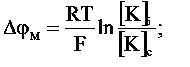
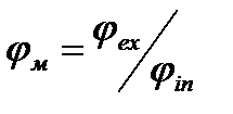
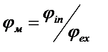
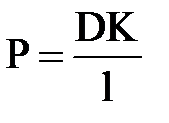
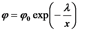
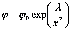
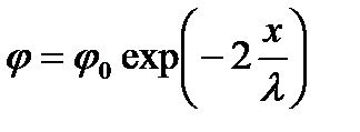
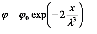
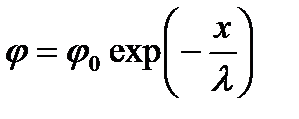
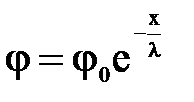
5. Обеспечивает через биопотенциалов 101. Biopotentials: 1. are potentials emerging in cells, tissues and organs in the process of their life activity; 2. electrical voltage emerging in spatial structural substances; 3. potential difference of two points of any conductor; 4. electric current emerging in living medium; 5. electric current emerging in spatial structural substances. 102. Registering of tissues and organs biopotentials: 1. autoradiography; 2. electrography; 3. X-ray diagnostics; 4. thermography; 5. phonocardiography. 103. Resting potential: 1. potential difference between cytoplasm of unexcited cell and environment; 2. potential of electric field within the unexcited cell and environment; 3. potential emerging on the internal side of unexcited cell membrane; 4. potential emerging on the external side of unexcited cell membrane; 5. potential of magnetic field within the unexcited cell and environment. 104. At excitation potential difference between a cell and environment: 1. action potential arises; 2. potential difference arises; 3. internal forces arise; 4. external forces arise; 5. forces potential arises. 105. Potential difference between cytoplasm and environment: 1. external forces; 2. internal forces; 3. resting potential; 4. action potential; 5. action force. 106. Equation of equilibrium membrane potential: 1. Poiseuille equation; 2. Nernst equation; 3. Newton equation; 4. Hagen equation; 5. Hooke’s law. 107. Nernst equation: 1. 2. 3. 4. 5. 108. Goldman equation: 1. 2. 3. 4. 5. 109. Formula of membrane permeability coefficient: 1. 2. 3. 4. 5. 110. Electric voltage arising in cells and tissues of biological objects: 1. electric field; 2. electromagnetic waves; 3. biopotentials; 4. biological membranes; 5. electroconductivity. 111. A process corresponding to the action potential: 1. magnetization; 2. demagnetization; 3. heat release; 4. depolarization and repolarization; 5. polarization. 112. Phases of action potential: 1. magnetization; 2. demagnetization; 3. heat release; 4. ascending and descending; 5. polarization. 113. Permeability of membrane at cell excitation in initial period: 1. increases for K+ ions; 2. decreases for Na+ ions; 3. decreases for K+ ions; 4. increases for Na+ ions; 5. increases for Cl- ions. 114. Action potential propagates along the nervous fiber without attenuation: 1. in air medium; 2. in inactive medium; 3. in active medium; 4. in isotropic medium; 5. in anisotropic medium. 115. Charge of intacellular medium in comparison with extracellular one: 1. in rest is negative, in maximum of action potential is positive; 2. in rest is positive, in maximum of action potential is negative; 3. is always positive; 4. is always negative; 5. always equals to zero. 116. Condition of the arising of action potential: 1. presence of potassium and sodium concentration gradients; 2. presence of chlorine ions concentration gradient; 3. excessive diffusion of magnesium ions; 4. excessive diffusion of calcium ions; 5. excessive diffusion of phosphorus ions. 117. Potentials of ionic type: 1. diffusive, membrane, phase; 2. diffusive, membrane, passive; 3. membrane, phase, active; 4. diffusive, membrane; 5. diffusive, membrane, resting potential. 118. Duration of cardiomyocyte action potential in comparison with the axon action potential: 1. more; 2. less; 3. the same; 4. equals to zero; 5. doesn’t change. 119. Plateau phase in cardiomyocytes is determined by ion fluxes: 1. JNa inside, JK inside; 2. JK inside, JCl inside; 3. JK outside, JCa inside; 4. JNa outside, JH+ inside; 5. JCa inside, JMg inside. 120. Depolarization phase in cardiomyocytes is determined by ion fluxes: 1. JNa inside; 2. JK inside; 3. JK outside; 4. JNa outside; 5. JCa inside. 121. Repolarization phase in cardiomyocytes is determined by ion fluxes: 1. JNa inside; 2. JK inside; 3. JK outside; 4. JNa outside; 5. JCa inside. 122. Ionic channels in biological membranes: 1. independently on ∆φм; 2. conductivity of channels depends on Т; 3. channel transfer K+, Na+ and Сa2+ the same way; 4. there are separate channels for different types of ions; 5. conductivity of channels depends on φ. 123. Resting potential: 1. corresponds to the repolarization process; 2. corresponds to the polarization process; 3. corresponds to the depolarization process; 4. corresponds to the refractoriness process; 5. corresponds to the refractoriness and depolarization processes. 124. At the resting state cytoplasmic membrane is maximally permeable for ions of: F) К G) Na H) Cl I) Ca J) Mg 125. Ascending phase of action potential: 1. corresponds to the repolarization process; 2. corresponds to the polarization process; 3. corresponds to the depolarization process; 4. corresponds to the refractoriness process; 5. corresponds to the refractoriness and depolarization processes. 126. Membrane potential φм: 1. 2. 3. 4. 5. 127. At the resting state ratio of membrane permeability coefficients of squid axon for different ions is: 1. Pk:РNa:Pcl=0.04:1:0.45 2. Pk:РNa:Pcl=1:20:0.45 3. Pk:РNa:Pcl=1:0.04:0.45 4. Pk:РNa:Pcl=20:0.04:0.45 5. Pk:РNa:Pcl=0.45:0.04:1 128. At the excitation state ratio of membrane permeability coefficients of squid axon for different ions is: 1. Pk:РNa:Pcl=0.04:1:0.45 2. Pk:РNa:Pcl=1:20:0.45 3. Pk:PNa:Pcl=1:0.04:0.45 4. Pk:РNa:Pcl=20:0.04:0.45 5. Pk:РNa:Pcl=0.45:0.04:1 129. Excitation of the membrane: 1. is described by Goldman equation; 2. is described by Newton equation; 3. is described by Hodgkin-Huxley equation; 4. is described by Nernst equation; 5. is described by Einstein equation. 130. Hodgkin-Huxley equation: 1. 2. 3. 4. 5. 131. Absolute value of equilibrium potential of Nernst: 1. doesn’t change with temperature increasing; 2. decreases with temperature increasing; 3. increases with temperature increasing; 4. initially increases then decreases with temperature increasing; 5. initially decreases then increases with temperature increasing; 132. Absolute value of Goldman-Hodgkin-Katz stationary potential: 1. initially increases then decreases with temperature increasing; 2. initially decreases then increases with temperature increasing; 3. doesn’t change with temperature increasing; 4. increases with temperature increasing; 5. decreases with temperature increasing. 133. Biopotentials are subdivided into: 1. equilibrium, nonequilibrium, simple. 2. active, passive, impulse; 3. muscular, neuro-cerebral, diffusive; 4. phasic, nonequilibrium, active; 5. diffusive, membrane, phasic. 134. Action potential arises at: 1. stationary state; 2. transfer of substances; 3. excitation, potential difference between the cell and environment; 4. excitation, temperature difference in membrane and cell; 5. membrane excitation. 135. General changing of potential on the membrane occurring at cell excitation: 1. density of substance flux through the membrane; 2. resting potential; 3. membrane potential; 4. distribution of potential in nervous fiber; 5. action potential. 136. In the moment pf excitation polarity of membrane changes to opposite: 1. polarization; 2. repolarization; 3. depolarization; 4. deformation; 5. reverberation. 137. Electrodes for biopotentials removal: 1. are used in ballistocardiography, mechanocardiography; 2. are used in phonocardiography, ultrasound diagnostics; 3. are used in encephalography, cardiography; 4. are used in ultrasound diagnostics rheography; 5. are used in mechanocardiography. 138. Founder of membrane theory of potentials: 1. Bernstein; 2. Einstein; 3. Röntgen; 4. Huxley; 5. Galvani. 139. First time experimentally measured the potential difference on the membrane of living cell: 1. Hodgkin-Huxley; 2. Einthoven; 3. Goldman; 4. Schrödinger; 5. Nernst-Planck. 140. A process that decreases negative potential within the cell: 1. depolarization; 2. repolarization; 3. polarization; 4. deformation; 5. reverberation. 141. Method of the registering the bioelectrical activity of a muscle: 1. encephalography; 2. electrography; 3. echoencephalography; 4. electromyography; 5. electrocardiography. 142. If in certain point of unmyelinated fiber the potential equaled to φ0 then at x distance from this point it will be: 1. 2. 3. 4. 5. 143. Nervous fibers: 1. myelinated and unmyelinated; 2. plasmatic and non-plasmatic; 3. excitated and unexcited; 4. actin; 5. myosin. 144. Excitation of some part of unmyelinated nervous fiber leads to: 1. local depolarization of the membrane; 2. transport of ions; 3. passive transport; 4. active transport; 5. hyperpolarization. 145. Telegrapher’s equation for nervous fibers: 1. 2. 3. 4. 5.
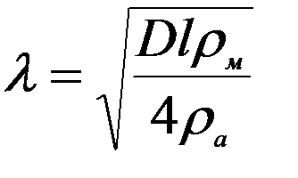
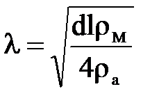
146. Constant length of a nervous fiber:
1. 2. 3. 4. 5. 147. Solution of the telegrapher’s equation: 1. 2. 3. 4. 5. E=gradU 148. In depolarization phase at axon excitation flows of Na+ ions are directed: 1. JNa inside the cell; 2. JNa out of the cell; 3. JNa=0 4. active; 5. passive. 149. In axon repolarization phase flows of ions are directed: 1. J Na inside the cell; 2. JК inside the cell; 3. JК out of the cell; 4. active; 5. passive. 150. Покое потенциала нервной клетки приближается к равновесному: potential of nervous cell approximates to the equilibrium: 1. calcium potential; 2. sodium potential; 3. chlorine potential; 4. potassium potential; 5. protons potential. 151. During the generation of action potential nervous cell potential approaching to the equilibrium: 1. calcium potential; 2. sodium potential; 3. chlorine potential; 4. potassium potential; 5. protons potential. 152. Propagation of action potential along the myelinated fiber: 1. continuous; 2. saltatory (intermittent); 3. constant; 4. alternating; 5. infinite. 153. Propagation of action potential along the unmyelinated fiber: 1. continuous; 2. saltatory (intermittent); 3. constant; 4. alternating; 5. infinite. 154. Special intercellular connections that are used to the passing of a signal from one cell to another is called: 1. neurotransmitter; 2. synapse; 3. action potential; 4. node of Ranvier; 5. Schwann cell. 155. The structure providing the passing of a signal from ending of axon of a nervous cell to a neuron, muscular fiber, secretory cell is called: 1. neurotransmitter; 2. synapse; 3. action potential; 4. node of Ranvier 5. Schwann cell. 156. Myelin sheath of nerve fiber of haemoglobin molecules: 1. consists of sphingosine molecules; 2. consists of protein-lipid complex; 3. consists of red blood cells molecules; 4. consists of calcium molecules. 157. During the dreaming delta-rhythm arises, 1. 0,5-3,5 Hz; till 300 µV; 2. 8-13 Hz; till 200 µV; 3. 8-13 Hz; till 300 µV; 4. 3,5-7,5 Hz; till 100 µV; 5. 15-100 Hz; till 100 µV. 158. Recording of biological processes (biopotentials, biocurrents) in the structure of brain is implemented by: 1. tomograph; 2. encephalograph; 3. phonocardiograph; 4. rheograph; 5. laser. 159. Myelin sheath that surround areas of nervous cells is kind of: 1. plasmatic membrane; 2. nervous fiber; 3. neurolemma; 4. sarcolemma; 5. karyolemma. 160. Conducts nervous impulses from the body of a cell and dendrites to other neurons: 1. synapse; 2. axon; 3. plasmatic reticulum; 4. soma; 5. neurilemma. 161. Extension of neuron (short) that transmits nervous impulses to the neuron body: 1. synapse; 2. axon; 3. plasmatic reticulum; 4. soma; 5. dendrite. 162. The great importance for EEG genesis has: 1. interrelation of electrical activity of pyramidal neurons; 2. interrelation between cerebral cortex and electrodes; 3. interrelation between electrodes; 4. totality of current electrical details of separate neurons; 5. arithmetic mean of the potential differences. 163. Model of cerebral cortex electrical activity: 1. Franck model; 2. Gibbs energy; 3. Einstein model; 4. Zhadin model; 5. Stoletov model. 164. Electroencephalography is: 1. method of registering of muscle bioelectrical activity; 2. method of registering biopotentials that arise in cardiac muscle at its excitation; 3. method of registering of brain bioelectrical activity; 4. method of measuring the heart sizes in dynamics; 5. method of measuring the blood flow velocity. 165. Main indexes of EEG value are: 1. frequency and amplitude of these oscillations; 2. changing of the potential difference; 3. changing of temperature difference; 4. standard deviation of these oscillations; 5. arithmetic mean of the potential differences. 166. Generation of exciting postsynaptic potential in the area of dendrite trunk without the branching leads to the emergence of: 1. quadrupole; 2. dendrite dipole; 3. action potential; 4. resting potential; 5. somatic dipole. 167. Taken from the surface of body biopotential is measured in: 1. milliampere; 2. millivolt; 3. nanometre; 4. micrometre; 5. centimetre. 168. Types of electrical activity of pyramidal neurons: 1. impulse and gradual potentials; 2. action potential; 3. resting potential; 4. resting potentials and interaction potentials; 5. interaction potentials. 169. Gradual (slow) potentials: 1. moving postsynaptic potentials; 2. inhibitory and excitatory postsynaptic potentials; 3. resting potential; 4. action potential; 5. transforming potentials. 170. Inhibitory postsynaptic potentials of pyramidal cells are generated: 1. in outer side of neurons; 2. между нейронами и головного мозга 3. in the body of neuron; 4. in inner side of neurons; 5. in dendrites. 171. Excitatory postsynaptic potentials of pyramidal cells are generated: 1. in outer side of neurons; 2. между нейронами и головного мозга 3. in the body of neuron; 4. in inner side of neurons; 5. in dendrites. 172. EEG genesis: 1. by gradual electrical activity of pyramidal neurons; 2. by impulse activity of pyramidal neurons; 3. by electrical activity of dipoles; 4. by electrical activity of cells; 5. by electrical activity of soma. 173. Changing of membrane potential of pyramidal neurons is explained: 1. by presence of alternating electric field; 2. by presence of direct electric field; 3. by presence of impulse current; 4. by presence of differing from each other somatic and dendritic dipoles; 5. by the changing of dipole moments. 174. Potential that is formed by somatic dipole: 1. inhibitory postsynaptic potential; 2. excitatory postsynaptic potential; 3. action potential; 4. resting potential; 5. membrane potential. 175. Potential that is formed by dendritic dipole: 1. inhibitory postsynaptic potential; 2. excitatory postsynaptic potential; 3. action potential; 4. resting potential; 5. membrane potential. 176. Direction of vector of dendritic dipole: 1. perpendicular to neurons; 2. parallel to neurons; 3. from soma along dendritic trunk; 4. towards soma along dendritic trunk; 5. from neurons to environment. 177. Direction of vector of somatic dipole: 1. perpendicular to neurons; 2. parallel to neurons; 3. from soma along dendritic trunk; 4. towards soma along dendritic trunk; 5. from neurons to environment. 178. EEG signals: 1. superultrasound; 2. powerful; 3. weak and powerful; 4. constant; 5. alternating and weak. 179. Distribution of neurons in the cortex of brain: 1. irregular and their dipole moments are perpendicular to the surface of cortex; 2. regular and their dipole moments are perpendicular to the surface of cortex; 3. irregular and their dipole moments are parallel to the surface of cortex; 4. regular and their dipole moments are parallel to the surface of cortex; 5. chaotically. 180. Bonds between activities of pyramidal neurons: 1. covalent; 2. strongly negative; 3. weakly negative; 4. positive correlation; 5. negative correlation. 181. Quantities characterizing EEG indexes: 1. amplitude and frequency of oscillations of potential difference; 2. impedance of electrical circuit; 3. direction of propagating oscillations; 4. wave velocity; 5. period of oscillations of potential difference. 182. In rest (at absence of irritators) EEG registers: 1. α-rhythm; 2. β-rhythm; 3. γ-rhythm; 4. δ-rhythm; 5. σ-rhythm. 183. At the active state of the brain EEG registers: 1. α-rhythm; 2. β-rhythm; 3. γ-rhythm; 4. δ-rhythm; 5. σ-rhythm. 184. During the sleeping EEG registers: 1. α-rhythm; 2. β-rhythm; 3. γ-rhythm; 4. δ-rhythm; 5. σ-rhythm. 185. At nerve excitation EEG registers: 1. α-rhythm; 2. β-rhythm; 3. γ-rhythm; 4. δ-rhythm; 5. σ-rhythm. 186. In rest (at absence of irritators) EEG registers α-rhythms with frequencies: 1. (8 - 13) Hz; 2. (0,5 - 3,5) Hz; 3. (14 - 30) Hz; 4. (30 - 55) Hz and higher; 5. higher than 100 Hz. 187. At the active state of the brain EEG registers β-rhythm with frequencies: 1. (8 - 13) Hz; 2. (0,5 - 3,5) Hz; 3. (14 - 30) Hz; 4. (30 - 55) Hz and higher; 5. higher than 100 Hz. 188. During the sleeping EEG registers δ-rhythm with frequencies: 1. (8 - 13) Hz; 2. (0,5 - 3,5) Hz; 3. (14 - 30) Hz; 4. (30 - 55) Hz and higher; 5. higher than 100 Hz. 189. At nerve excitation EEG registers γ-rhythm with frequencies: 1. (8 - 13) Hz; 2. (0,5 - 3,5) Hz; 3. (14 - 30) Hz; 4. (30 - 55) Hz and higher; 5. higher than 100 Hz. 190. Electroencephalography is: 1. registering and analysis of brain biopotentials; 2. registering and analysis of heart biopotentials; 3. registering and analysis of skin biopotentials; 4. registering and analysis of eye retina biopotentials; 5. registering and analysis of nerve trunks and muscles biopotentials. 191. Method of research the mechanical indexes of heart work: 1. ballistocardiography; 2. phonocardiography; 3. echocardiography; 4. electrocardiography; 5. encephalography. 192. Echocardiography is the method of research the structure and movement of heart structures with usage of: 1. alternating current of high frequency; 2. Compton effect; 3. absorbed X-ray radiation; 4. reflected ultrasound; 5. impedance registering. 193. Registering of time dependence of heart biopotentials in electrocardiograph is implemented with: 1. amplifier; 2. source of calibration voltage; 3. electrodes; 4. ultrasound generator; 5. condenser. 194. Electrocardiography is: 1. method of registering of muscle bioelectrical activity at its excitation; 2. method of registering biopotentials that arise in cardiac muscle at its excitation; 3. method of registering of brain bioelectrical activity; 4. method of measuring the heart sizes in dynamics; 5. method of measuring the blood flow velocity. 195. Electrodes applied on a patient at electrography are targeted for taking the: 1. electrical moment of heart; 2. current between two points on body surface; 3. potential difference between two points on body surface; 4. charges formed by heart on body surface; 5. magnetic moment of heart. 196. Problems of the research of electric fields in an organism: 1. determination the electrical resistance of tissues and organs; 2. studying the changing of electrical impulses form; 3. studying the influence of environment to the emergence of electrical potentials; 4. diagnostics of diseases; 5. registering of organs and tissues biopotentials in norm and pathology for the diagnosis of a disease. 197. Electromyography: 1. method of registering the bioelectrical activity of muscles; 2. method of registering the biopotentials arising in cardiac muscle at its excitation; 3. method of registering the bioelectrical activity of brain; 4. method of measuring the heart sizes in dynamics; 5. method of measuring the blood flow velocity. 198. Vector of electric moment of dipole characterizing heart biopotentials: 1. electric vector of polarization; 2. strength of electric field of dipole; 3. strength of magnetic field of dipole; 4. integral electric vector; 5. Umov-Poynting vector. 199. Main characteristic of dipole: 1. impulse moment; 2. electric moment; 3. moment of force; 4. moment of inertia; 5. velocity gradient. 200. On the base of registering the time dependence of heart magnetic field induction method of…is created: 1. electrocardiography; 2. electromyography; 3. electroradiography; 4. ballistocardiography; 5. magnetocardiography. 201. Different disorders of heart functioning that lead to the violation of normal heart rate: 1. extrasystoly; 2. stenocardia; 3. atherosclerosis; 4. thrombophlebitis; 5. arrythmia. 202. Difference of amplitudes of the same ECG prongs in the same moment of time in different leads: 1. the value of integral electrical vector E is different for different leads; 2. rotation of the vector E is different in different leads; 3. projections of the vector E to the different leads are not the same; 4. for each lead there is its own vector E; 5. projections of the vector E to the different leads are the same.
|




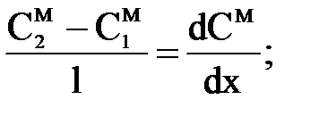


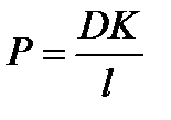 ;
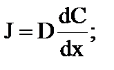
;
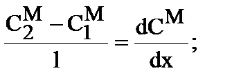


 ;
;
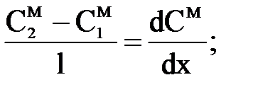


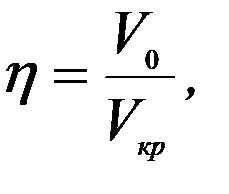


 - slow high amplitude oscillations of electrical activity of brain. Specify the diapason: